2017-11-17
Six hours for healing
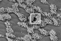
Scanning electron microscope image of airway epithelium with damaged cells (withe box) and clustered cilia on some cells. (Source: Kretschmer et al., 2017, Copyright American Society for Investigative Pathology)
In a recent publication in the American Journal of Pathology, DZL researchers from Lübeck University describe for the first time in which way small injuries of the airway epithelium heal. Thus, these wounds close in the time frame of six hours. The researchers damaged tissue areas with a laser beam and observed subsequent repair process by a combination of microscopic techniques.
The integrity of the airway epithelium is vitally important. Only intact tissue can maintain physiological lung function. Moreover, the epithelium is the first barrier against invading pathogens. What happens, if it is damaged? How long does it take until a wound is closed and which mechanisms are important?
In order to answer these questions, scientists of Lübeck University retrieved tissue samples from mouse trachea. Using a focused laser beam, they damaged small tissue areas (1-12 cells) and observed the repair process by different microscopic techniques. They found that wounds of a size up to six cells close within six hours. Only two hours after the laser impulse the protein F actin – it initiates cell movement – was detected. Later, the surrounding cells protrude into the wounded area and push destroyed out of the wounds (see figure). Finally, the cellular debris is conveyed out of the airways by cilia. Larger wounds didn’t close during the observation period of six hours. In many of these cases, the basement membrane was damaged. As observation was limited to shorter time periods, it remains elusive, if similar repair mechanisms apply for larger wounds, too.
With these experiments, the scientists from Lübeck advanced their tissue investigation technique based on observation of autofluorescence. This microscopic method, described last year in another paper, enables them to follow dynamic processes in explanted tissues. “Therefore, our latest findings are a technological advance as well as biologically relevant”, says Professor Peter König, deputy head of the University’s Institute of Anatomy. Tissue repair is one of DZL’s overarching topics as it is of importance in a number of lung diseases. The experiments presented here have been performed on trachea of mice which are regarded as a model for small human airways. Future investigations shall elucidate, if these repair processes also occur in the human lung.
Further Information: Kretschmer S, Pieper M, Klinger A, Hüttmann G, König P (2017) Imaging of Wound Closure of Small Epithelial Lesions in the Mouse Trachea. Am J Pathol 187: 2451-2460
This article was published open access. The DZL supports open access publications, as they are freely available for everyone. There is no restriction by subscription of the respective journal. Thus, new scientific knowledge is proliferating faster and distributed without constraints.
/jbul
Calendar
Upcoming events
2024-04-30
Virtual DZL AID Kick-off Seminar
2024-05-29
74th Meeting of ARCN
2024-06-05